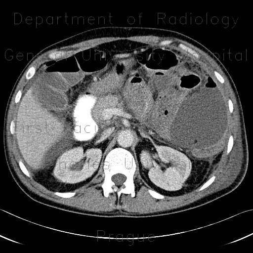
Crohn's disease of small bowel, abdominal abscess, CT routine and CT enterography

CASE

Patient with known Crohn's disease with involvement of aboral ileum and a segment in the mid-part of the small bowell. The extent of the disease was first recognized on CT enterography (last two images). Later, the patient presented with acute abdomen and a routine CT with iodine peroral contrast was performed, which showed formation of gigantic thin-walled abscess with an air-fluid level, most likely due to perforation of the bowel.

Radiology image - Crohn

Crohn's disease of small bowel, abdominal abscess


Crohn's disease of small bowel, abdominal abscess: CT - Computed tomographyCrohn's disease of small bowel, abdominal abscess: CT - Computed tomographyCrohn's disease of small bowel, abdominal abscess: CT - Computed tomographyCrohn's disease of small bowel, abdominal abscess: CT - Computed tomographyCrohn's disease of small bowel, abdominal abscess: CT - Computed tomographyCrohn's disease of small bowel, abdominal abscess: CT - Computed tomographyCrohn's disease of small bowel, abdominal abscess: CT - Computed tomographyCrohn's disease of small bowel, abdominal abscess, CT enterography: CT - Computed tomographyCrohn's disease of small bowel, abdominal abscess, CT enterography: CT - Computed tomography
lights on off