Crohn's disease, stenosis, prestenotic dilatation, rupture, CT enteroclysis, enterography

CASE

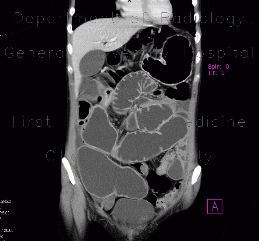
Patient with Crohn's disease had several strictures and signs of prestenotic dilatation at baseline CT enteroclysis examination. After three years, he developed rupture of the small intestine, which was treated with acute surgery. Follow-up examination shows persistent stenotic segments with chronic prestenotic dilatation.

Radiology image - Crohn

Crohn's disease, stenosis, prestenotic dilatation, CT enteroclysis, first examination


Crohn's disease, stenosis, prestenotic dilatation, CT enteroclysis, first examination: CT - Computed tomographyCrohn's disease, stenosis, prestenotic dilatation, CT enteroclysis, first examination: CT - Computed tomographyCrohn's disease, stenosis, rupture, routine CT at 3 years: CT - Computed tomographyCrohn's disease, stenosis, rupture, routine CT at 3 years: CT - Computed tomographyCrohn's disease, stenosis, rupture, routine CT at 3 years: CT - Computed tomographyCrohn's disease, stenosis, rupture, routine CT at 3 years: CT - Computed tomographyCrohn's disease, stenosis, prestenotic dilatation, CT enterography at 3 years: CT - Computed tomographyCrohn's disease, stenosis, prestenotic dilatation, CT enterography at 3 years: CT - Computed tomographyCrohn's disease, stenosis, prestenotic dilatation, CT enterography at 3 years: CT - Computed tomography
lights on off