ATLAS OF RADIOLOGICAL IMAGES v.1
General University Hospital and 1st Faculty of Medicine of Charles University in Prague
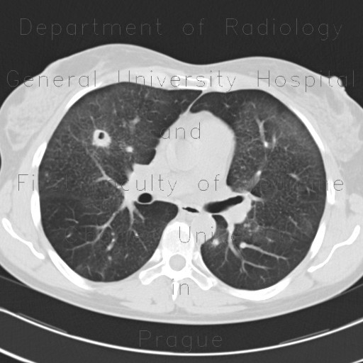
Wegener's granulomatosis, development in time
CASE
A sequence of CT scans shows development of pulmonary Wegener's granulomatosis in time, in five years. It shows enlargement of one cavitated mass in the left lower lung lobe with an air-fluid level in the second year, extensive relapse in the third year with formation of a large mass in the left lung lobe, which gradually diminished in time.


