ATLAS OF RADIOLOGICAL IMAGES v.1
General University Hospital and 1st Faculty of Medicine of Charles University in Prague
Acute pancreatitis, chylous pancreatitis, hypertriglyceridemia-associated acute pancreatitis
CASE
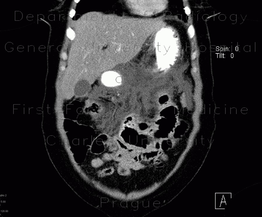
CT images of a patient with hypertriglyceridemia-associated pancreatitis show wast areas of fat necrosis in the peripancreatic fat, mesocolon transversum, and fluid on both prerenal fascias, around the liver. The pancreas itself is enlarged but enhances homogeneously.
CT images of a patient with hypertriglyceridemia-associated pancreatitis show wast areas of fat necrosis in the peripancreatic fat, mesocolon transversum, and fluid on both prerenal fascias, around the liver. The pancreas itself is enlarged but enhances homogeneously.">


