ATLAS OF RADIOLOGICAL IMAGES v.1
General University Hospital and 1st Faculty of Medicine of Charles University in Prague
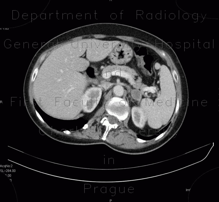
Adrenal metastasis and skeletal metastasis of pulmonary carcinoma, correlation
CASE
This case shows correlation between CT, bone scintigraphy and ultrasound. CT shows a soft-tissue mass in the mediastinum and the right hilum. Ultrasound and CT show an oval osteolytic metastasis of the ninth left rib, that has a correlation with increased uptake on bone scintigraphy. It is hypoechoic on ultrasound and has enhancing periphery on CT. Bilateral adrenal metastases have enhancing periphery and necrotic center. They are hypoechoic on ultrasound. Note also pericardial and pleural effusion.


