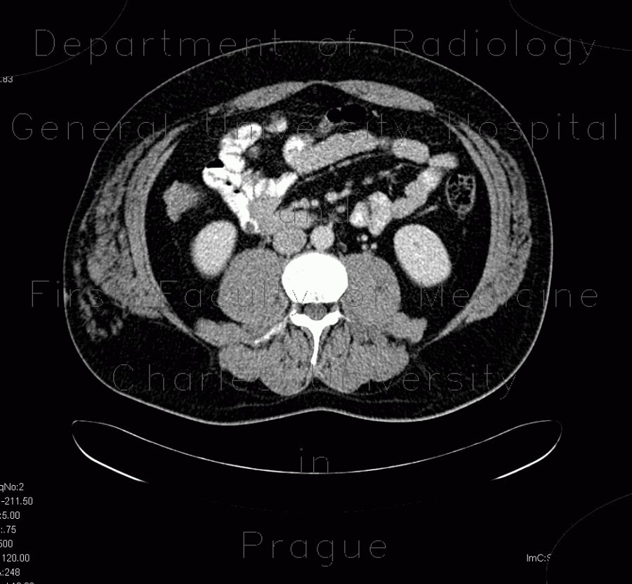
Angiolipoma, subcutaneous, selective embolisation

CASE

CT shows a mass of fat density with interlacing lacelike soft tissue structures, which represent vessels, and calcifications in the vessels. Ultrasound shows a mass that is isoechogeneous to the subcutaneous tissue and contains several vessels that are seen on colour map. This lesion was treated by selective embolisation.

Radiology image - Angiolipoma, subcutaneous, selective embolisation: Abdomen, Soft tissue, Vessels: CT - Computed tomography

Angiolipoma, subcutaneous, selective embolisation


Angiolipoma, subcutaneous, selective embolisation: CT - Computed tomographyAngiolipoma, subcutaneous, selective embolisation: CT - Computed tomographyAngiolipoma, subcutaneous, selective embolisation: CT - Computed tomographyAngiolipoma, subcutaneous, selective embolisation: CT - Computed tomographyAngiolipoma, subcutaneous, selective embolisation: CT - Computed tomographyAngiolipoma, subcutaneous, selective embolisation: CT - Computed tomographyAngiolipoma, subcutaneous, selective embolisation: CT - Computed tomographyAngiolipoma, subcutaneous, selective embolisation: CT - Computed tomographyAngiolipoma, subcutaneous, selective embolisation: CT - Computed tomographyAngiolipoma, subcutaneous, selective embolisation: CT - Computed tomographyAngiolipoma, subcutaneous, selective embolisation: CT - Computed tomographyAngiolipoma, subcutaneous, selective embolisation: US - UltrasoundAngiolipoma, subcutaneous, selective embolisation: US - UltrasoundAngiolipoma, subcutaneous, selective embolisation: US - UltrasoundAngiolipoma, subcutaneous, selective embolisation: AG - AngiographyAngiolipoma, subcutaneous, selective embolisation: AG - AngiographyAngiolipoma, subcutaneous, selective embolisation: AG - AngiographyAngiolipoma, subcutaneous, selective embolisation: AG - AngiographyAngiolipoma, subcutaneous, selective embolisation: AG - AngiographyAngiolipoma, subcutaneous, selective embolisation: AG - Angiography
lights on off