ATLAS OF RADIOLOGICAL IMAGES v.1
General University Hospital and 1st Faculty of Medicine of Charles University in Prague
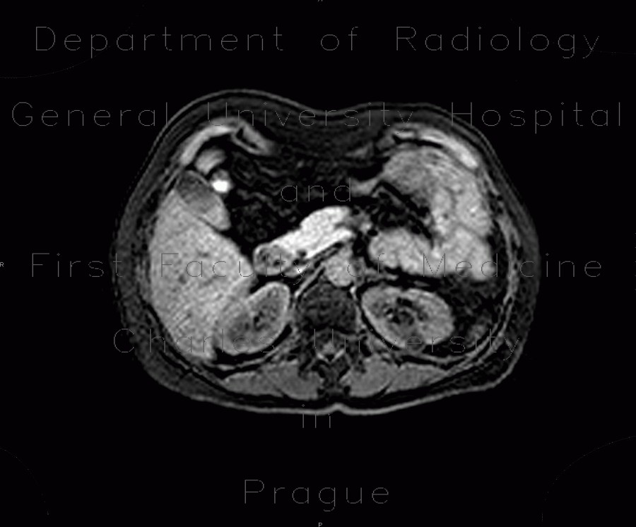
Angiomyolipoma of kidney, atypical
CASE
CT shows a small tumour arising from the left cortex of the left kidney. It has somewhat increased density. Ultrasound showed that the nodule has increased echogenicity and is homogeneous and well-defined. MRI study concluded, that a presence of atypical angiomyolipoma (with limited fat content) is more likely.


