ATLAS OF RADIOLOGICAL IMAGES v.1
General University Hospital and 1st Faculty of Medicine of Charles University in Prague
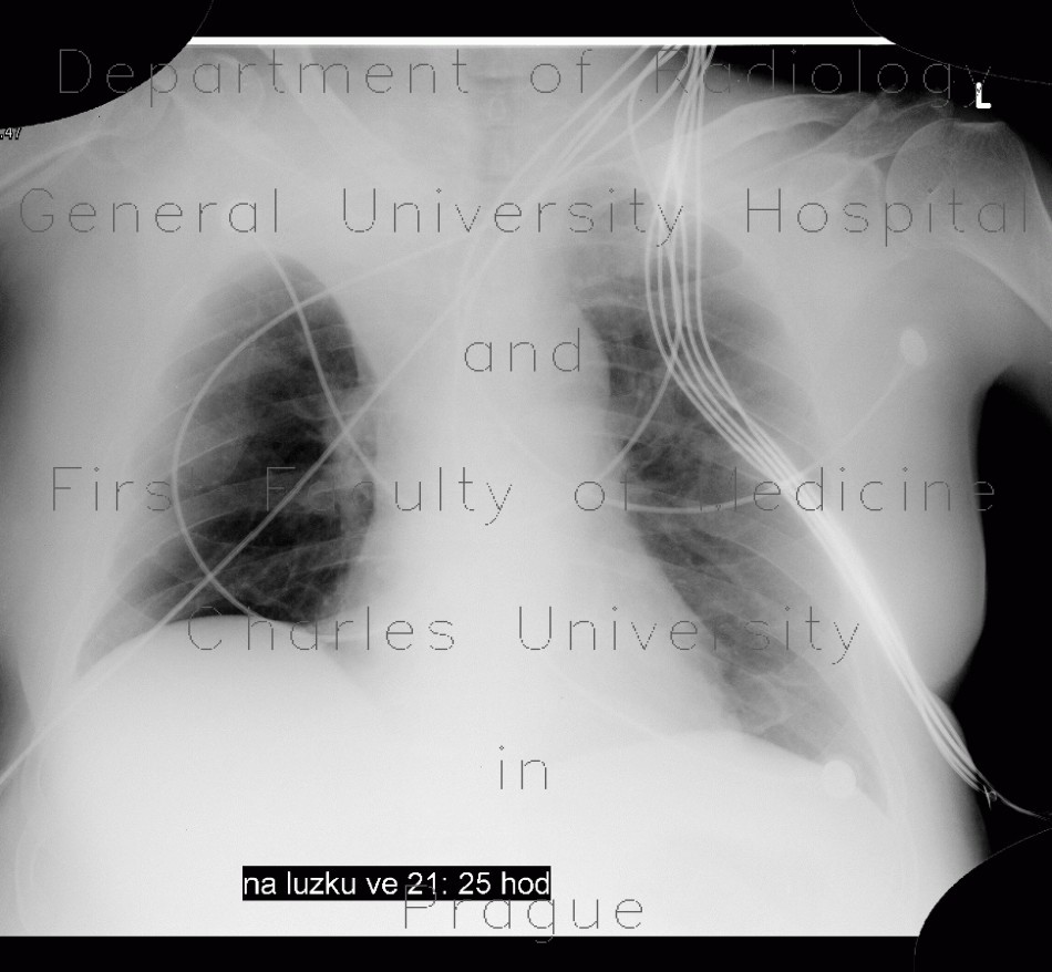
Atelectasis of right upper lobe of lung
CASE
AP supine chest radiograph shows thin area of decreased transparency along the right border of upper mediastinum and the apex which is sharply delianted by its concave border. Also, the paratracheal stripe is effaced and right upper lobe vascular structures are obscured. CT shows decreased aeration of the right upper bronchi due to obstruction of the lobar bronchus and collapse of the right upper lobe. The third chest radiograph shows, that the collapse was ultimately resolved.


