ATLAS OF RADIOLOGICAL IMAGES v.1
General University Hospital and 1st Faculty of Medicine of Charles University in Prague
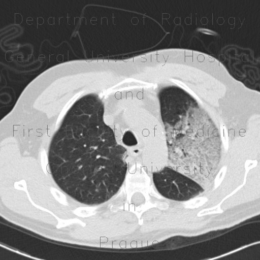
Atypical pneumonia, influenza, H1N1, 10 months after
CASE
CT shows areas of ground-glass and septal thickening in lobar and lobular distribution. This adult male patient was diagnosed with H1N1 influenza strain. Ten months after, he underwent follow-up HRCT showing fibrous bands and residual areas of consolidation in subpleural region.


