ATLAS OF RADIOLOGICAL IMAGES v.1
General University Hospital and 1st Faculty of Medicine of Charles University in Prague
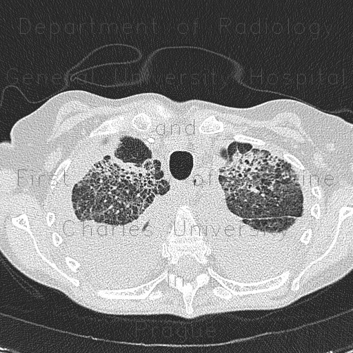
Atypical pneumonia, subacute stage, lung fibrosis
CASE
In subacute stage of atypical pneumonia (Pneumocystis carinii), an image almost indistinguishable from fibrosis may develop. This patient developed bilateral pleural effusion, extensive areas of septal thickening and distortion of parechymal architecture with bronchiectasis.


