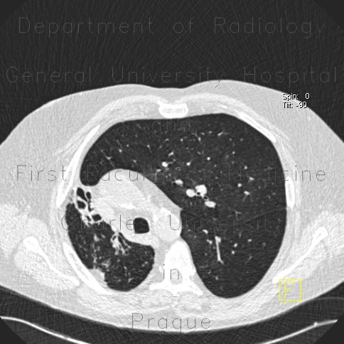
Bronchiectasias, postspecific changes

CASE

CT shows decreased volume of the right hemithorax with shift of mediastinum towards the right side and elevation of the hemidiphragm. Lower and middle lobes are replaced by dilated bronchi, which are seen also in the right upper lunge lobe. These changes are attributed do tuberculosis in the past.

Tags:  Thorax  Lung
Radiology image - Bronchiectasias, postspecific changes: Thorax, Lung: CT - Computed tomography

Bronchiectasias, postspecific changes


Bronchiectasias, postspecific changes: CT - Computed tomographyBronchiectasias, postspecific changes: CT - Computed tomographyBronchiectasias, postspecific changes: CT - Computed tomographyBronchiectasias, postspecific changes: CT - Computed tomographyBronchiectasias, postspecific changes: CT - Computed tomographyBronchiectasias, postspecific changes: CT - Computed tomographyBronchiectasias, postspecific changes: CT - Computed tomography
lights on off