Bronchopneumonia, biopsy, recurrence

CASE

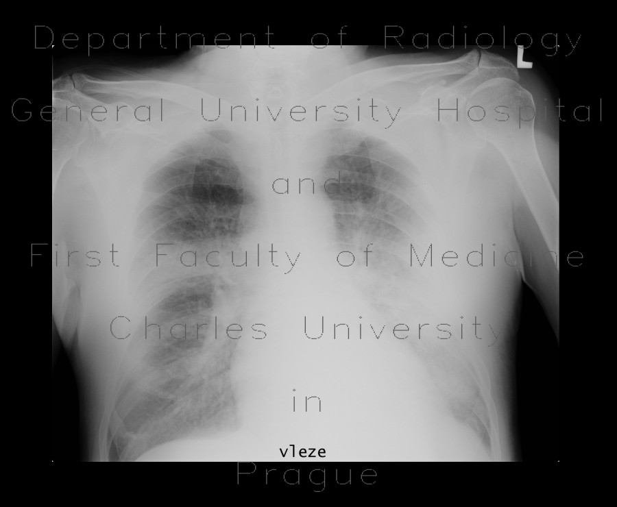
Plain radiographs show infiltrative shadows in both lung wings. Subsequent CT shows airspace consolidation consistent with acute bronchopneumonia, which was confirmed by a biopsy requested by the physician. Follow-up CT shows regression of bronchopneumonia infiltrates leaving a small area of fibrous changes. Unfortunately, next CT showed recurrence of bronchopneumonia.

Tags:  Thorax  Lung
Radiology image - Bronchopneumonia, biopsy: Thorax, Lung: X-ray - Plain radiograph

Bronchopneumonia, biopsy


Bronchopneumonia, biopsy: X-ray - Plain radiographBronchopneumonia, biopsy: X-ray - Plain radiographBronchopneumonia, biopsy: CT - Computed tomographyBronchopneumonia, biopsy: CT - Computed tomographyBronchopneumonia, biopsy: CT - Computed tomographyBronchopneumonia, biopsy: CT - Computed tomographyBronchopneumonia, biopsy: CT - Computed tomographyBronchopneumonia, biopsy: CT - Computed tomographyBronchopneumonia, biopsy, follow-up: CT - Computed tomographyBronchopneumonia, biopsy, follow-up: CT - Computed tomographyBronchopneumonia, biopsy, recurrence: CT - Computed tomographyBronchopneumonia, biopsy, recurrence: CT - Computed tomographyBronchopneumonia, biopsy, recurrence: CT - Computed tomographyBronchopneumonia, biopsy, recurrence: CT - Computed tomographyBronchopneumonia, biopsy, recurrence: CT - Computed tomographyBronchopneumonia, biopsy, recurrence: CT - Computed tomography
lights on off