ATLAS OF RADIOLOGICAL IMAGES v.1
General University Hospital and 1st Faculty of Medicine of Charles University in Prague
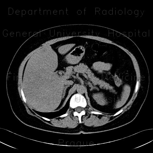
Carcinoma of sigmoid colon, metastasis in liver
CASE
CT shows multiple liver metastases with a typical appearance of hypodense center surrounded by hyperdense rim. CT also confirmed tumorous thickening of sigmoid colon. Initially, the ultrasound image of hypoechoic lesions in the liver parenchyma was mistaken for liver cysts and segmental thickening of sigmoid colon for diverticulitis. Liver metastases may appear on ultrasound almost anechoic and therefore mimic cysts, if the liver is steatotic as was in this case.


