ATLAS OF RADIOLOGICAL IMAGES v.1
General University Hospital and 1st Faculty of Medicine of Charles University in Prague
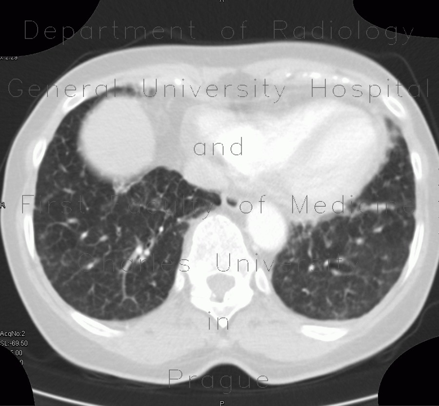
Carcinomatous lymphangoitis, lymphangoitis carcinosa, osteolysis of shoulder blade
CASE
Initial chest radiograph shows a portcatheter inserted in the superior vena cava. In comparison, the follow-up images show marked increase in lung markings especially in the right lower lobe due to thickened interlobular and axial interstitium. Note also osteolytic changes of the left shoulder blade.


