Cerebral hemorrhage, hypertonic hemorrhage, hemocephalus, subfalcine herniation, descendent transtentorial herniation

CASE

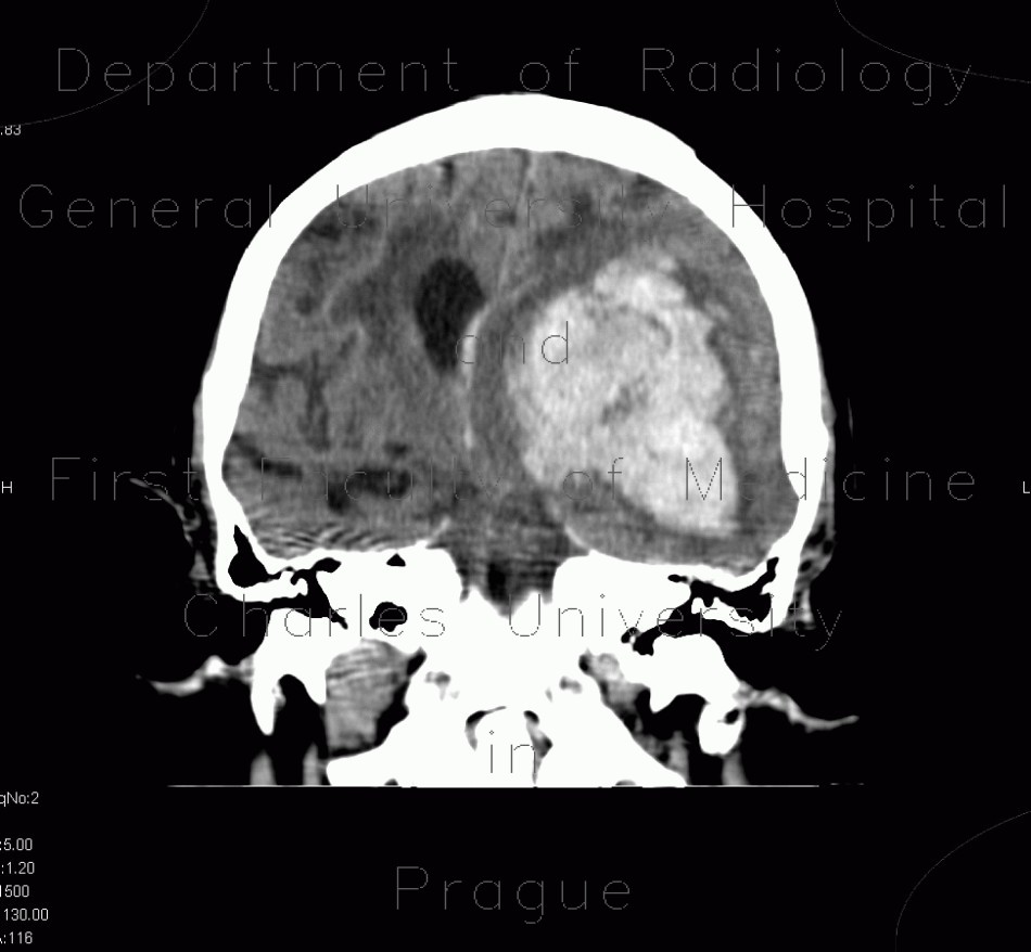
CT shows massive bleeding into the left frontal and parietal lobe which extended into the left and right lateral ventricles causing hemocephalus. The hemorrhage is surrounded by hypodense rim of edema and causes subfalcine herniation with severe shift of midline structures to the right. Note also that the left ambient cistern is obliterated which is a sign of transtentorial herniation.
Tags:  Brain  Brain
Radiology image - Cerebral hemorrhage, hypertonic hemorrhage, hemocephalus, subfalcine herniation, descendent transtentorial herniation: Brain, Brain: CT - Computed tomography

Cerebral hemorrhage, hypertonic hemorrhage, hemocephalus, subfalcine herniation, descendent transtentorial herniation


Cerebral hemorrhage, hypertonic hemorrhage, hemocephalus, subfalcine herniation, descendent transtentorial herniation: CT - Computed tomographyCerebral hemorrhage, hypertonic hemorrhage, hemocephalus, subfalcine herniation, descendent transtentorial herniation: CT - Computed tomographyCerebral hemorrhage, hypertonic hemorrhage, hemocephalus, subfalcine herniation, descendent transtentorial herniation: CT - Computed tomographyCerebral hemorrhage, hypertonic hemorrhage, hemocephalus, subfalcine herniation, descendent transtentorial herniation: CT - Computed tomographyCerebral hemorrhage, hypertonic hemorrhage, hemocephalus, subfalcine herniation, descendent transtentorial herniation: CT - Computed tomographyCerebral hemorrhage, hypertonic hemorrhage, hemocephalus, subfalcine herniation, descendent transtentorial herniation: CT - Computed tomography
lights on off