Cerebral ischemia, craniotomy, subarachnoid hemorrhage

CASE

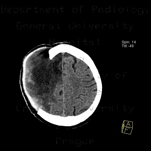
Extensive edema of right frontal, parietal, and temopral lobe following cerebral ischemia. Cerebral tissue herniates through the craniotomy opening, which was created for this purpose. Stripes of blood in the subarachnoid space and focus of intracerebral hemorrhage.

Tags:  Brain  Brain
Radiology image - Cerebral ischemia, craniotomy, subarachnoid hemorrhage: Brain, Brain: CT - Computed tomography

Cerebral ischemia, craniotomy, subarachnoid hemorrhage


Cerebral ischemia, craniotomy, subarachnoid hemorrhage: CT - Computed tomographyCerebral ischemia, craniotomy, subarachnoid hemorrhage: CT - Computed tomographyCerebral ischemia, craniotomy, subarachnoid hemorrhage: CT - Computed tomographyCerebral ischemia, craniotomy, subarachnoid hemorrhage: CT - Computed tomographyCerebral ischemia, craniotomy, subarachnoid hemorrhage: CT - Computed tomographyCerebral ischemia, craniotomy, subarachnoid hemorrhage: CT - Computed tomography
lights on off