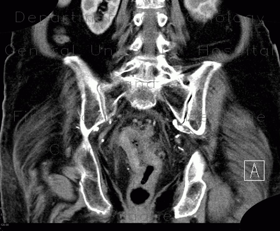
Colorectal cancer, tumorous stenosis of sigmoid colon, placement of stent

CASE

CT shows a relatively long segment of tumorous wall thickening in the rectosigmoid junction. Fluoroscopy shows a typical apple-core appearance of the tumorous stenosis. A stent was placed to ensure patency of this segment.

Radiology image - Colorectal cancer, tumorous stenosis of sigmoid colon, placement of stent: Abdomen, Large bowel: CT - Computed tomography

Colorectal cancer, tumorous stenosis of sigmoid colon, placement of stent


Colorectal cancer, tumorous stenosis of sigmoid colon, placement of stent: CT - Computed tomographyColorectal cancer, tumorous stenosis of sigmoid colon, placement of stent: CT - Computed tomographyColorectal cancer, tumorous stenosis of sigmoid colon, placement of stent: CT - Computed tomographyColorectal cancer, tumorous stenosis of sigmoid colon, placement of stent: CT - Computed tomographyColorectal cancer, tumorous stenosis of sigmoid colon, placement of stent: RF - FluoroscopyColorectal cancer, tumorous stenosis of sigmoid colon, placement of stent: RF - FluoroscopyColorectal cancer, tumorous stenosis of sigmoid colon, placement of stent: RF - FluoroscopyColorectal cancer, tumorous stenosis of sigmoid colon, placement of stent: RF - FluoroscopyColorectal cancer, tumorous stenosis of sigmoid colon, placement of stent: RF - FluoroscopyColorectal cancer, tumorous stenosis of sigmoid colon, placement of stent: RF - FluoroscopyColorectal cancer, tumorous stenosis of sigmoid colon, placement of stent: RF - FluoroscopyColorectal cancer, tumorous stenosis of sigmoid colon, placement of stent: RF - Fluoroscopy
lights on off