Contusion of frontopolar cortex, beam hardening artifact, subarachonid hemorrhage

CASE

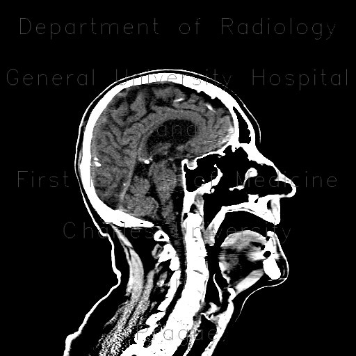
A patient, who sustained contusion of the skull with subcutaneous hematoma in the parietooccipital region, underwent CT of brain. Initial scan showed several dense foci in both frontopolar regions. This finding always has to be carefully interpreted, as in this patient, apart from beam-hardening artifact also small contusion foci were present. On follow-up examination after 24 hours, a full-blown image of contusions can be appreciated. Note also development of subarachonid hemorrhage around the occipital lobe.
Tags:  Brain  Brain
Radiology image - Contusion of frontopolar cortex, beam hardening artifact, subarachonid hemorrhage, initial scan: Brain, Brain: CT - Computed tomography

Contusion of frontopolar cortex, beam hardening artifact, subarachonid hemorrhage, initial scan


Contusion of frontopolar cortex, beam hardening artifact, subarachonid hemorrhage, initial scan: CT - Computed tomographyContusion of frontopolar cortex, beam hardening artifact, subarachonid hemorrhage, follow-up: CT - Computed tomographyContusion of frontopolar cortex, beam hardening artifact, subarachonid hemorrhage, initial scan: CT - Computed tomographyContusion of frontopolar cortex, beam hardening artifact, subarachonid hemorrhage, initial scan: CT - Computed tomographyContusion of frontopolar cortex, beam hardening artifact, subarachonid hemorrhage, follow-up: CT - Computed tomographyContusion of frontopolar cortex, beam hardening artifact, subarachonid hemorrhage, initial scan: CT - Computed tomographyContusion of frontopolar cortex, beam hardening artifact, subarachonid hemorrhage, follow-up: CT - Computed tomography
lights on off