Diaphragmatic hernia, Morgagni hernia

CASE

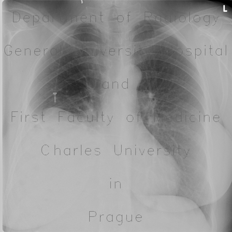
Plain chest radiographs shows marked elevation of the left hemidiaphragm. CT shows, that it is caused by a right-sided diaphragmatic hernia which contains right liver lobe, gallbladder with gallstones, and large bowel. It causes compressive changes of the right lowe lung lobe. This hernia was treated surgically with a good result which can be appreciated on the last two CT images.

Radiology image - Diaphragmatic hernia, Morgagni hernia: Abdomen, Biliary tree, Large bowel, Liver, Lung, Soft tissue: X-ray - Plain radiograph

Diaphragmatic hernia, Morgagni hernia


Diaphragmatic hernia, Morgagni hernia: X-ray - Plain radiographDiaphragmatic hernia, Morgagni hernia: X-ray - Plain radiographDiaphragmatic hernia, Morgagni hernia: CT - Computed tomographyDiaphragmatic hernia, Morgagni hernia: CT - Computed tomographyDiaphragmatic hernia, Morgagni hernia: CT - Computed tomographyDiaphragmatic hernia, Morgagni hernia: CT - Computed tomographyDiaphragmatic hernia, Morgagni hernia: CT - Computed tomographyDiaphragmatic hernia, Morgagni hernia: CT - Computed tomographyDiaphragmatic hernia, Morgagni hernia: CT - Computed tomographyDiaphragmatic hernia, Morgagni hernia: CT - Computed tomographyDiaphragmatic hernia, Morgagni hernia: CT - Computed tomographyDiaphragmatic hernia, Morgagni hernia: CT - Computed tomography
lights on off