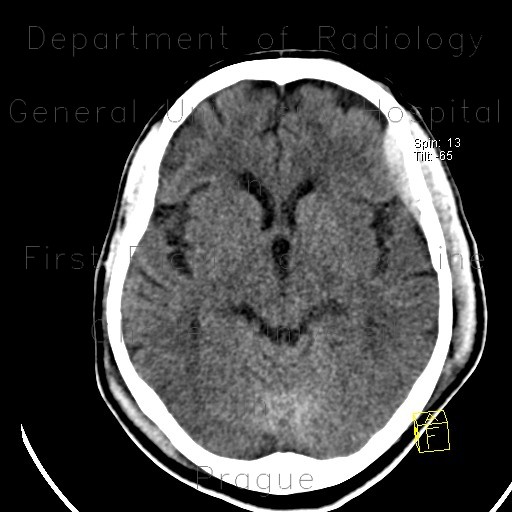
Epidural hematoma, subarachoid hemorrhage, cerebral contusion, skull fissure

CASE

A hairline fissure traverses left part of coronary suture. Adjacent to it, there is a lenticular circumscribed hyperdensity abutting the inner aspect of the skull, a typical epidural hematoma. Thin hairline densities interdigiating with sulci represent subarachnoid hemorrhage. Small cerebral contusion with perifocal oedema in the proximity.

Radiology image - Epidural hematoma, subarachoid hemorrhage, cerebral contusion, skull fissure: Brain, Head and Neck, Bone, Brain: CT - Computed tomography

Epidural hematoma, subarachoid hemorrhage, cerebral contusion, skull fissure


Epidural hematoma, subarachoid hemorrhage, cerebral contusion, skull fissure: CT - Computed tomographyEpidural hematoma, subarachoid hemorrhage, cerebral contusion, skull fissure: CT - Computed tomographyEpidural hematoma, subarachoid hemorrhage, cerebral contusion, skull fissure: CT - Computed tomographyEpidural hematoma, subarachoid hemorrhage, cerebral contusion, skull fissure: CT - Computed tomographyEpidural hematoma, subarachoid hemorrhage, cerebral contusion, skull fissure: CT - Computed tomographyEpidural hematoma, subarachoid hemorrhage, cerebral contusion, skull fissure: CT - Computed tomography
lights on off