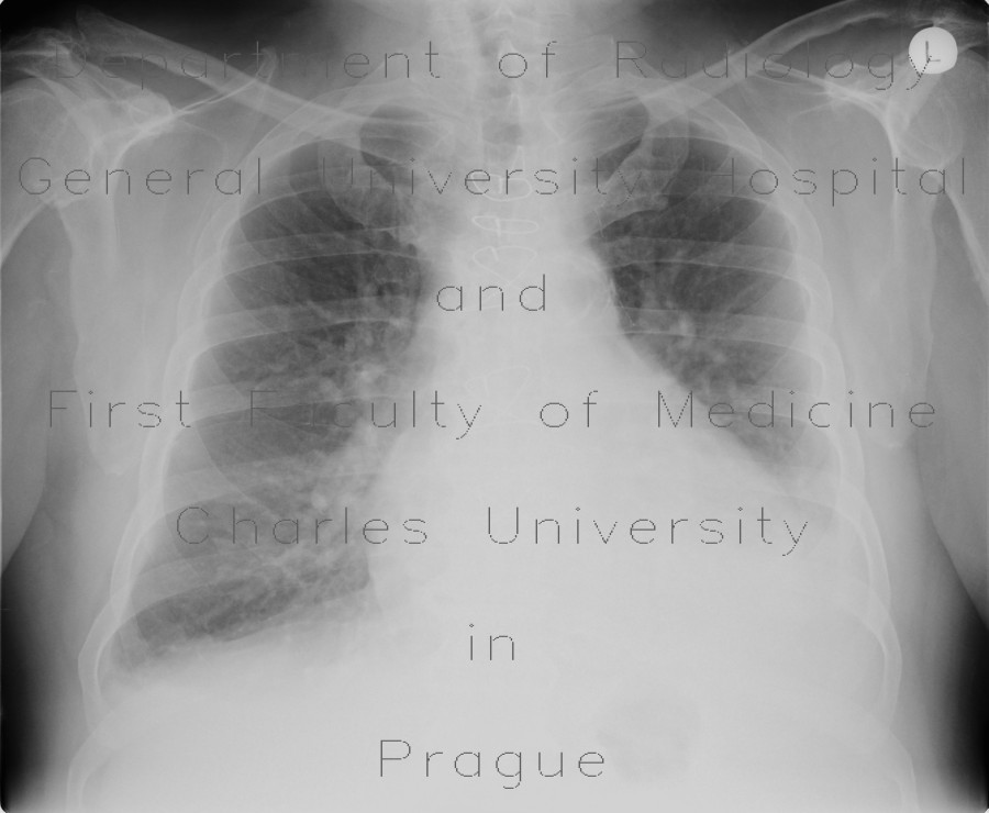
Fluidothorax, hemothorax and pneumothorax, complication of evacuation

CASE

Initially, there was some amount of pleural fluid in the left pleural cavity. An attempt was made to evacuate this fluid. It resulted in hemothorax seen as markedly decreased transparency of the left hemithorax on supine chest radiograph. Pneumothorax with collapse of the left upper lung lobe is shown on the third radiograph, where a chest tube is already in place. CT shows massive amount of fluid in the right pleural cavity. This fluid has increased density indicating hemothorax.

Radiology image - Fluidothorax, hemothorax and pneumothorax, complication of evacuation: Thorax, Lung, Mediastinum and pleural cavity: X-ray - Plain radiograph

Fluidothorax, hemothorax and pneumothorax, complication of evacuation


Fluidothorax, hemothorax and pneumothorax, complication of evacuation: X-ray - Plain radiographFluidothorax, hemothorax and pneumothorax, complication of evacuation: X-ray - Plain radiographFluidothorax, hemothorax and pneumothorax, complication of evacuation: X-ray - Plain radiographFluidothorax, hemothorax and pneumothorax, complication of evacuation: CT - Computed tomographyFluidothorax, hemothorax and pneumothorax, complication of evacuation: CT - Computed tomographyFluidothorax, hemothorax and pneumothorax, complication of evacuation: CT - Computed tomographyFluidothorax, hemothorax and pneumothorax, complication of evacuation: CT - Computed tomographyFluidothorax, hemothorax and pneumothorax, complication of evacuation: CT - Computed tomography
lights on off