Fluidothorax, pneumothorax, pneumomediastinum, subcutaneous emphysema, muscular emphysema, pneumocolum

CASE

Marked sucutaneous and muscular emphysema, small pneumothorax on both sides ventrally, fluidothorax and compression of dorsobasal parts of both lung wings. A chest tube in the left hemithorax positioned to the apex.
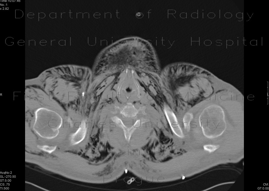
Radiology image - Fluidopneumothorax, pneumomediastinum, subcutaneous emphysema, muscular emphysema, pneumocolum: Thorax, Lung, Mediastinum and pleural cavity: CT - Computed tomography

Fluidopneumothorax, pneumomediastinum, subcutaneous emphysema, muscular emphysema, pneumocolum


Fluidopneumothorax, pneumomediastinum, subcutaneous emphysema, muscular emphysema, pneumocolum: CT - Computed tomographyFluidopneumothorax, pneumomediastinum, subcutaneous emphysema, muscular emphysema, pneumocolum: CT - Computed tomographyFluidopneumothorax, pneumomediastinum, subcutaneous emphysema, muscular emphysema, pneumocolum: CT - Computed tomographyFluidopneumothorax, pneumomediastinum, subcutaneous emphysema, muscular emphysema, pneumocolum: CT - Computed tomographyFluidopneumothorax, pneumomediastinum, subcutaneous emphysema, muscular emphysema, pneumocolum: CT - Computed tomography
lights on off