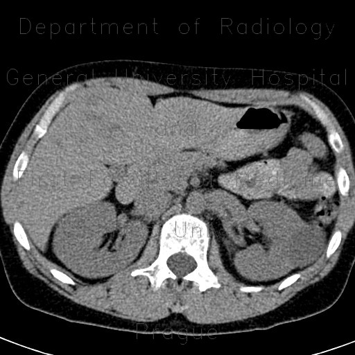
Focal nodular hyperplasia, FNH, flow rate of contrast

CASE

A large mass in the right liver lobe with central hypodensity shows only slightly increased enhancement in the arterial phase compared to surrounding parenchyma which is caused by low flow rate of contrast.

Tags:  Abdomen  Liver
Radiology image - Focal nodular hyperplasia, FNH, flow rate of contrast: Abdomen, Liver: CT - Computed tomography

Focal nodular hyperplasia, FNH, flow rate of contrast


Focal nodular hyperplasia, FNH, flow rate of contrast: CT - Computed tomographyFocal nodular hyperplasia, FNH, flow rate of contrast: CT - Computed tomographyFocal nodular hyperplasia, FNH, flow rate of contrast: CT - Computed tomographyFocal nodular hyperplasia, FNH, flow rate of contrast: CT - Computed tomographyFocal nodular hyperplasia, FNH, flow rate of contrast: CT - Computed tomographyFocal nodular hyperplasia, FNH, flow rate of contrast: CT - Computed tomographyFocal nodular hyperplasia, FNH, flow rate of contrast: CT - Computed tomographyFocal nodular hyperplasia, FNH, flow rate of contrast: CT - Computed tomographyFocal nodular hyperplasia, FNH, flow rate of contrast: CT - Computed tomography
lights on off