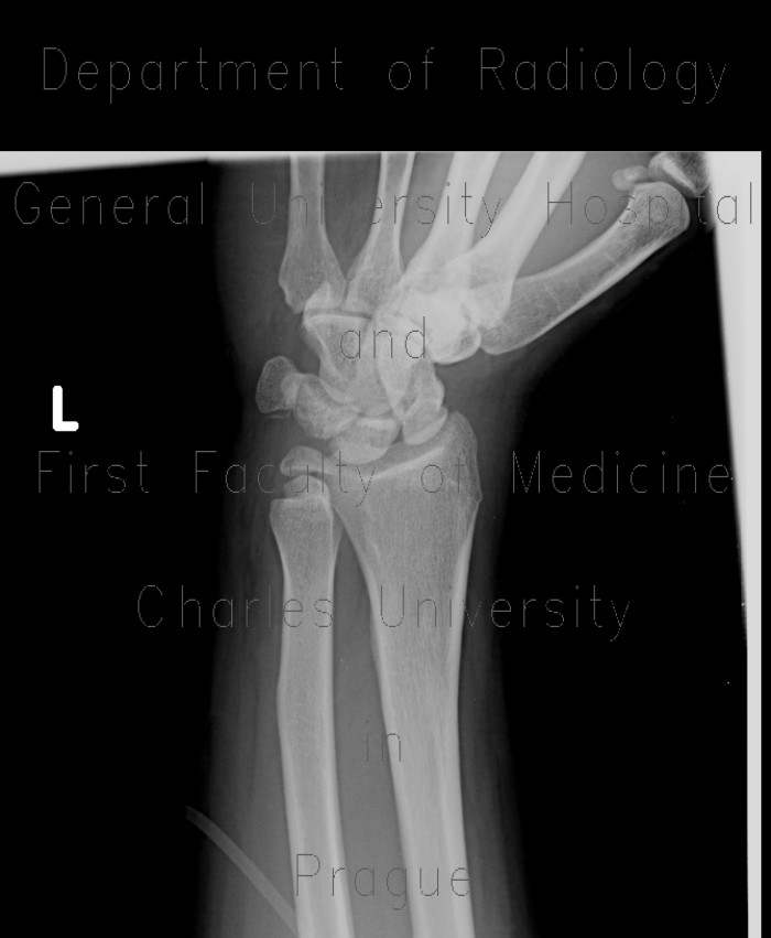
Fracture of scaphoid bone, dislocation of pisiform bone, abruption of lunate

CASE

Fracture of the waist of scaphoid bone. Abruption of small fragment of volar edge of lunate, which is rotated and dislocated palmarly. The pisiform bone is shifted to the ulnar side.

Tags:  Extremity  Bone
Radiology image - Fracture of scaphoid bone, dislocation of pisiform bone, abruption of lunate: Extremity, Bone: X-ray - Plain radiograph

Fracture of scaphoid bone, dislocation of pisiform bone, abruption of lunate


Fracture of scaphoid bone, dislocation of pisiform bone, abruption of lunate: X-ray - Plain radiographFracture of scaphoid bone, dislocation of pisiform bone, abruption of lunate: X-ray - Plain radiographFracture of scaphoid bone, dislocation of pisiform bone, abruption of lunate: X-ray - Plain radiographFracture of scaphoid bone, dislocation of pisiform bone, abruption of lunate: X-ray - Plain radiographFracture of scaphoid bone, dislocation of pisiform bone, abruption of lunate: CT - Computed tomographyFracture of scaphoid bone, dislocation of pisiform bone, abruption of lunate: CT - Computed tomographyFracture of scaphoid bone, dislocation of pisiform bone, abruption of lunate: CT - Computed tomographyFracture of scaphoid bone, dislocation of pisiform bone, abruption of lunate: CT - Computed tomographyFracture of scaphoid bone, dislocation of pisiform bone, abruption of lunate: CT - Computed tomographyFracture of scaphoid bone, dislocation of pisiform bone, abruption of lunate: CT - Computed tomographyFracture of scaphoid bone, dislocation of pisiform bone, abruption of lunate: CT - Computed tomographyFracture of scaphoid bone, dislocation of pisiform bone, abruption of lunate: CT - Computed tomography
lights on off