ATLAS OF RADIOLOGICAL IMAGES v.1
General University Hospital and 1st Faculty of Medicine of Charles University in Prague
Hemangioma, atypical hemangioma
CASE
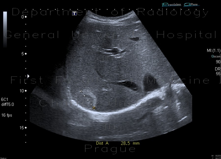
Ultrasound images shows a round subcapsular lesion in the right liver lobe. The lesion is isoechoic to liver parenchyma, outlined by a thick hyperechoic rim. The lesion exhibits posterior enhancement, which makes it likely to be an atypical hemangioma.


