Hemangioma of liver, cavernous hemangioma, capillary hemangioma

CASE

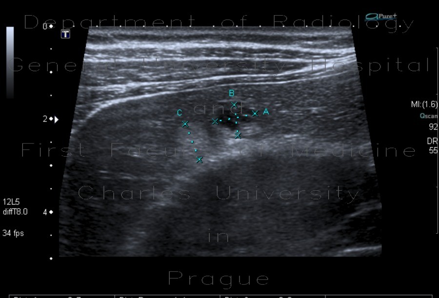
Ultrasound reveals two lesions in liver parenchyma - the first is hypoechoic, the second is hyperechoic, both are well-defined, with posterior acoustic enhancement. Color image show vascularisation in the first lesion, but nothing in the latter. The first lesion represents cavernous hemangioma and the second capillary hemangioma. Cavernous hemangiomas have larger vascular spaces that allow depiction of flow on ultrasound.

Tags:  Abdomen  Liver
Radiology image - Hemangioma of liver, cavernous hemangioma, capillary hemangioma: Abdomen, Liver: US - Ultrasound

Hemangioma of liver, cavernous hemangioma, capillary hemangioma


Hemangioma of liver, cavernous hemangioma, capillary hemangioma: US - UltrasoundHemangioma of liver, cavernous hemangioma, capillary hemangioma: US - UltrasoundHemangioma of liver, cavernous hemangioma, capillary hemangioma: US - UltrasoundHemangioma of liver, cavernous hemangioma, capillary hemangioma: US - UltrasoundHemangioma of liver, cavernous hemangioma, capillary hemangioma: US - UltrasoundHemangioma of liver, cavernous hemangioma, capillary hemangioma: US - UltrasoundHemangioma of liver, cavernous hemangioma, capillary hemangioma: US - UltrasoundHemangioma of liver, cavernous hemangioma, capillary hemangioma: US - Ultrasound
lights on off