ATLAS OF RADIOLOGICAL IMAGES v.1
General University Hospital and 1st Faculty of Medicine of Charles University in Prague
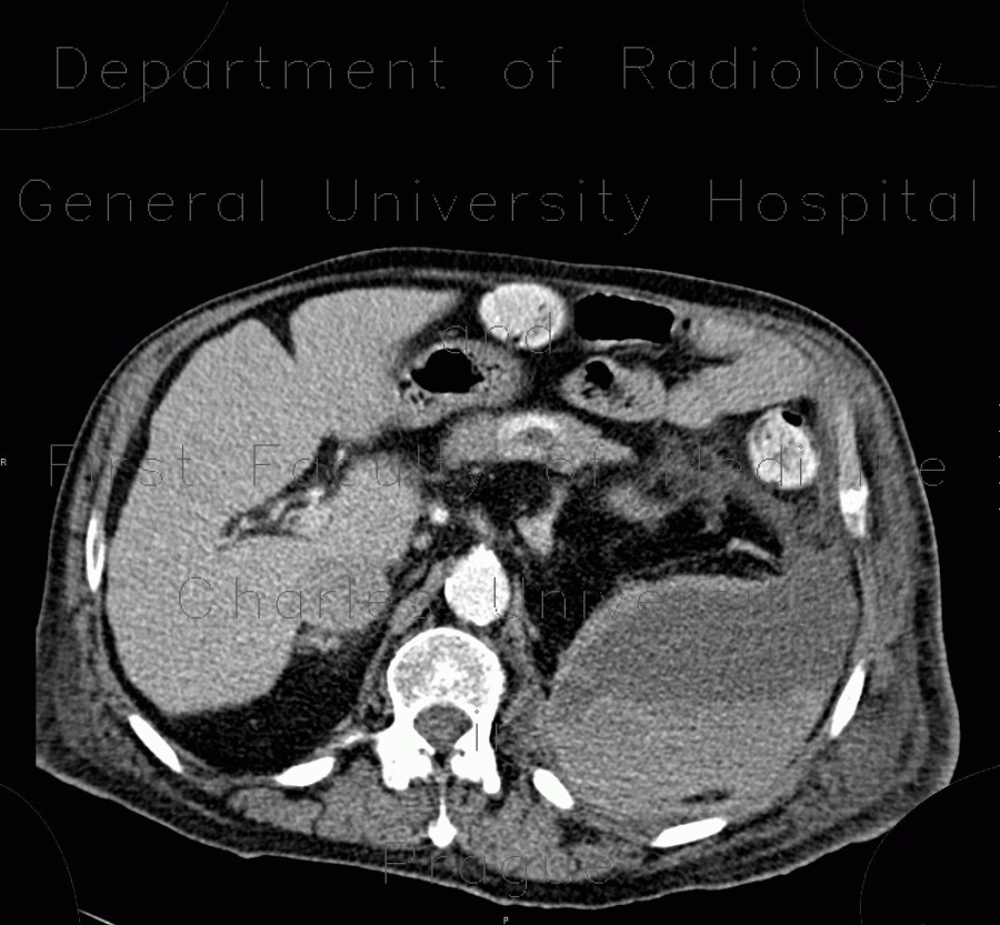
Hematoma in psoas muscle, large, anticoagulation therapy, anticoagulant
CASE
CT shows a large expansive mass involving left half of retroperitoneal space including psoas muscle. It is heterogeneous due to sedimentation of blood due to anticoagulant use. The thin hyperdense middle layer represents active bleeding of blood enhanced with intravenous contrast.


