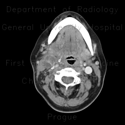
Inflammatory lymph node with liquefaction

CASE

CT shows an enlarged upper jugular node on the right side that contains necrotic hypodense areas with intervening normal enhancing cortex. Ultrasound shows an enlarged hypoechoic node with areas of effaced structure, increased polar vascularity and mild edema of surrounding fat, which has somewhat increased echogenicity.

Radiology image - Inflammatory lymph node with liquefaction: Head and Neck, Lymphatic: CT - Computed tomography

Inflammatory lymph node with liquefaction


Inflammatory lymph node with liquefaction: CT - Computed tomographyInflammatory lymph node with liquefaction: CT - Computed tomographyInflammatory lymph node with liquefaction: CT - Computed tomographyInflammatory lymph node with liquefaction: CT - Computed tomographyInflammatory lymph node with liquefaction: CT - Computed tomographyInflammatory lymph node with liquefaction: CT - Computed tomographyInflammatory lymph node with liquefaction: US - UltrasoundInflammatory lymph node with liquefaction: US - UltrasoundInflammatory lymph node with liquefaction: US - Ultrasound
lights on off