Inflammatory tumour of descending colon, CT colonography

CASE

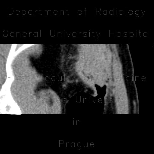
There is a mass withing the lumen of the sigmoid colon that protrudes into the aboral part and thus creating an appearance of a claw. This segment is surrounded by mesenteric fat of slightly increased density due to edema and mild dilatation of vasa recta. There is also mild reaction in the adjacent abdominal wall.

Radiology image - Inflammatory tumour of descending colon, CT colonography: Abdomen, Large bowel: CT - Computed tomography

Inflammatory tumour of descending colon, CT colonography


Inflammatory tumour of descending colon, CT colonography: CT - Computed tomographyInflammatory tumour of descending colon, CT colonography: CT - Computed tomographyInflammatory tumour of descending colon, CT colonography: CT - Computed tomographyInflammatory tumour of descending colon, CT colonography: CT - Computed tomographyInflammatory tumour of descending colon, CT colonography: CT - Computed tomographyInflammatory tumour of descending colon, CT colonography: CT - Computed tomographyInflammatory tumour of descending colon, CT colonography: CT - Computed tomographyInflammatory tumour of descending colon, CT colonography: CT - Computed tomographyInflammatory tumour of descending colon, CT colonography: CT - Computed tomographyInflammatory tumour of descending colon, CT colonography: CT - Computed tomographyInflammatory tumour of descending colon, CT colonography: CT - Computed tomographyInflammatory tumour of descending colon, CT colonography: CT - Computed tomographyInflammatory tumour of descending colon, CT colonography: CT - Computed tomographyInflammatory tumour of descending colon, CT colonography: CT - Computed tomography
lights on off