ATLAS OF RADIOLOGICAL IMAGES v.1
General University Hospital and 1st Faculty of Medicine of Charles University in Prague
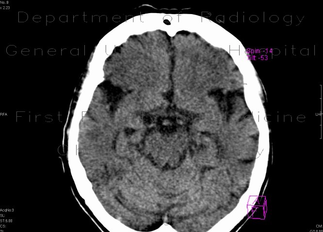
Ischemia, temporal lobe, development in time
CASE
Initially, only a discrete hint of hypodensity in the temporal lobe and flattening of gyri due to oedema. On the second follow-up, the development of ischemia is obvious - marked hypodensity in subcortical white matter and somewhat less in cortex. On the third CT there is apparent loss of corticomedullary diferentiation, the whole area is uniformely hypodense.


