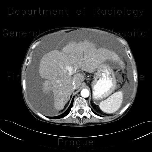
Liver cirrhosis, macronodular, ascites, massive

CASE

Both CT and ultrasound show cirrhotic liver with massive amount of ascites that delineates also peritoneal folds (mesentery, broad ligament of uterus, ...). Liver contours are undulated, and there is a loss of hyperechoic liver outline on ultrasound.

Radiology image - Liver cirrhosis, macronodular, ascites, massive: Abdomen, Liver, Peritoneal cavity: CT - Computed tomography

Liver cirrhosis, macronodular, ascites, massive


Liver cirrhosis, macronodular, ascites, massive: CT - Computed tomographyLiver cirrhosis, macronodular, ascites, massive: CT - Computed tomographyLiver cirrhosis, macronodular, ascites, massive: CT - Computed tomographyLiver cirrhosis, macronodular, ascites, massive: CT - Computed tomographyLiver cirrhosis, macronodular, ascites, massive: CT - Computed tomographyLiver cirrhosis, macronodular, ascites, massive: CT - Computed tomographyLiver cirrhosis, macronodular, ascites, massive: CT - Computed tomographyLiver cirrhosis, macronodular, ascites, massive: CT - Computed tomographyLiver cirrhosis, macronodular, ascites, massive: US - UltrasoundLiver cirrhosis, macronodular, ascites, massive: US - UltrasoundLiver cirrhosis, macronodular, ascites, massive: US - UltrasoundLiver cirrhosis, macronodular, ascites, massive: US - UltrasoundLiver cirrhosis, macronodular, ascites, massive: US - Ultrasound
lights on off