Lung contusion, hemothorax, pneumothorax, chest tube

CASE

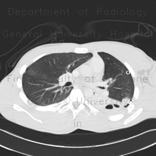
CT shows increased density of the left lung wing and less of the right lung wing. The ground glass opacity is most marked at the anterior margin, where it is with contact with chest wall, that was compressed, when the trauma occured. There is a small anterior pneumothorax on the left and blood (fluid of increased density) in the dependent position in the left pleural space. A chest tube was inserted into the left pleural space.

Radiology image - Lung contusion, hemothorax, pneumothorax, chest tube: Thorax, Lung, Mediastinum and pleural cavity: CT - Computed tomography

Lung contusion, hemothorax, pneumothorax, chest tube


Lung contusion, hemothorax, pneumothorax, chest tube: CT - Computed tomographyLung contusion, hemothorax, pneumothorax, chest tube: CT - Computed tomographyLung contusion, hemothorax, pneumothorax, chest tube: CT - Computed tomographyLung contusion, hemothorax, pneumothorax, chest tube: CT - Computed tomographyLung contusion, hemothorax, pneumothorax, chest tube: CT - Computed tomographyLung contusion, hemothorax, pneumothorax, chest tube: CT - Computed tomographyLung contusion, hemothorax, pneumothorax, chest tube: CT - Computed tomographyLung contusion, hemothorax, pneumothorax, chest tube: CT - Computed tomography
lights on off