ATLAS OF RADIOLOGICAL IMAGES v.1
General University Hospital and 1st Faculty of Medicine of Charles University in Prague
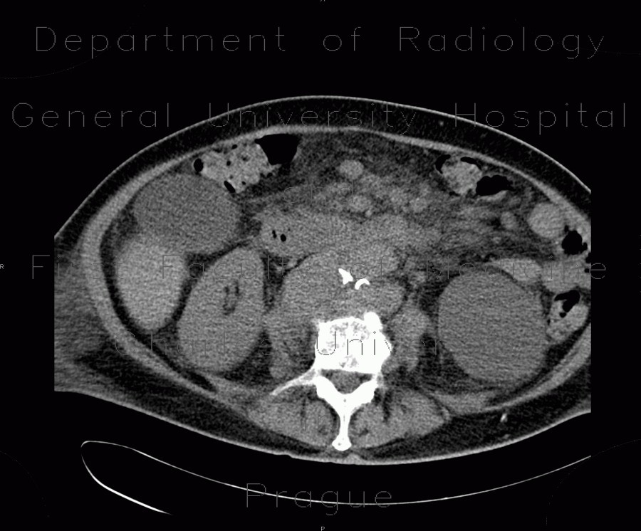
Lymphoma, mesenterial lymphadenopathy, edema of mesentery, correlation
CASE
This case shows correlation of CT and ultrasound images of mesenterial lymphadenopathy in a patient with lymphoma. CT shows multiple enlarged lymph nodes and increased density of the mesenteric fat. On ultrasound, the enlarged lymph nodes are hypoechoic and the edematous mesenteric fat is hyperechoic.


