ATLAS OF RADIOLOGICAL IMAGES v.1
General University Hospital and 1st Faculty of Medicine of Charles University in Prague
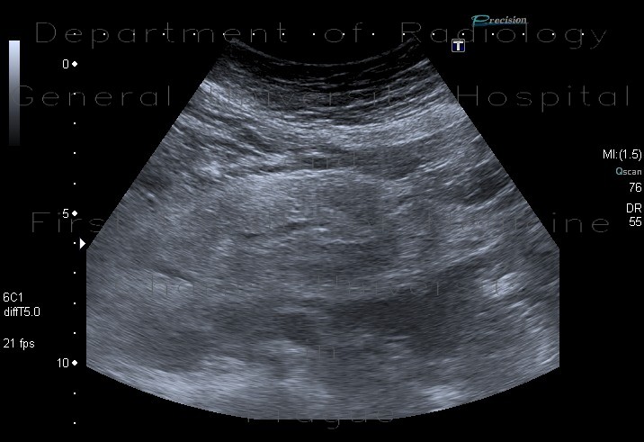
Mesenteric lipodystrophy, paniculitis
CASE
Ultrasound shows increased echogenicity of mesenteric fat. Several oval lymph nodes are barely seen due to obesity. The mesenteric fat has mass-like apearance and mesenteric vessels can be seen in its middle (hypoechoic linear structure in the first image).


