Multiple myeloma, venooclusive disease of liver, hypervascular nodules

CASE

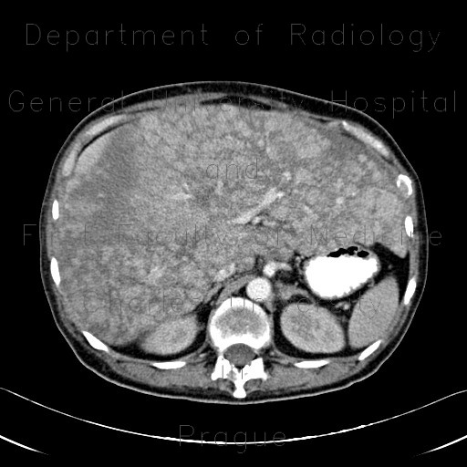
Patient with multiple myeloma with previously normal liver on ultrasound. The liver is markedly enlarged, show multiple nodules of increased enhancement that correspond to hypervascular nodules in venooclusive liver disease due to obstruction of small perisinusoidal branches of the hepatic veins. Multiple osteolytic lesions in bones.

Radiology image - Multiple myeloma, venooclusive disease of liver, hypervascular nodules: Abdomen, Spine and Axial, Bone, Liver: CT - Computed tomography

Multiple myeloma, venooclusive disease of liver, hypervascular nodules


Multiple myeloma, venooclusive disease of liver, hypervascular nodules: CT - Computed tomographyMultiple myeloma, venooclusive disease of liver, hypervascular nodules: CT - Computed tomographyMultiple myeloma, venooclusive disease of liver, hypervascular nodules: CT - Computed tomographyMultiple myeloma, venooclusive disease of liver, hypervascular nodules: CT - Computed tomographyMultiple myeloma, venooclusive disease of liver, hypervascular nodules: CT - Computed tomographyMultiple myeloma, venooclusive disease of liver, hypervascular nodules: CT - Computed tomographyMultiple myeloma, venooclusive disease of liver, hypervascular nodules: CT - Computed tomography
lights on off