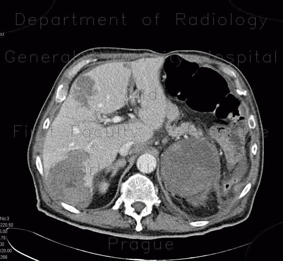
Neuroendocrine tumour of adrenal gland, liver metastasis, necrotic

CASE

CT shows a large mass in the left adrenal gland, which has heterogeneous structure with slightly enhancing soft tissue and necrotic component. There are hypodense confuent masses in the liver that represent necrotic metastases, which are heterogeneously hypoechoic on ultrasound.

Radiology image - Neuroendocrine tumour of adrenal gland, liver metastasis, necrotic: Abdomen, Kidney and adrenals, Liver: CT - Computed tomography

Neuroendocrine tumour of adrenal gland, liver metastasis, necrotic


Neuroendocrine tumour of adrenal gland, liver metastasis, necrotic: CT - Computed tomographyNeuroendocrine tumour of adrenal gland, liver metastasis, necrotic: CT - Computed tomographyNeuroendocrine tumour of adrenal gland, liver metastasis, necrotic: CT - Computed tomographyNeuroendocrine tumour of adrenal gland, liver metastasis, necrotic: CT - Computed tomographyNeuroendocrine tumour of adrenal gland, liver metastasis, necrotic: CT - Computed tomographyNeuroendocrine tumour of adrenal gland, liver metastasis, necrotic: CT - Computed tomographyNeuroendocrine tumour of adrenal gland, liver metastasis, necrotic: CT - Computed tomographyNeuroendocrine tumour of adrenal gland, liver metastasis, necrotic: CT - Computed tomographyNeuroendocrine tumour of adrenal gland, liver metastasis, necrotic: CT - Computed tomographyNeuroendocrine tumour of adrenal gland, liver metastasis, necrotic: US - UltrasoundNeuroendocrine tumour of adrenal gland, liver metastasis, necrotic: US - UltrasoundNeuroendocrine tumour of adrenal gland, liver metastasis, necrotic: US - Ultrasound
lights on off