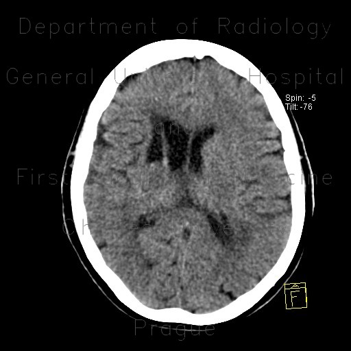
Occlusion of middle cerebral artery, MCA, collateral flow, postischemic changes

CASE

Both CT and angiography show, that the right middle cerebral artery is occluded at its origin (M1), however some collateral flow is still visible. Hypodense caput of the nucleus caudatus as a result of past ischemia.

Radiology image - Occlusion of middle cerebral artery, MCA, collateral flow, postischemic changes: Head and Neck, Brain, Vessels: CT - Computed tomography

Occlusion of middle cerebral artery, MCA, collateral flow, postischemic changes


Occlusion of middle cerebral artery, MCA, collateral flow, postischemic changes: CT - Computed tomographyOcclusion of middle cerebral artery, MCA, collateral flow, postischemic changes: CT - Computed tomographyOcclusion of middle cerebral artery, MCA, collateral flow, postischemic changes: CT - Computed tomographyOcclusion of middle cerebral artery, MCA, collateral flow, postischemic changes: CT - Computed tomographyOcclusion of middle cerebral artery, MCA, collateral flow, postischemic changes: CT - Computed tomographyOcclusion of middle cerebral artery, MCA, collateral flow, postischemic changes: CT - Computed tomographyOcclusion of middle cerebral artery, MCA, collateral flow, postischemic changes: CT - Computed tomographyOcclusion of middle cerebral artery, MCA, collateral flow, postischemic changes: CT - Computed tomographyOcclusion of middle cerebral artery, MCA, collateral flow, postischemic changes: CT - Computed tomographyOcclusion of middle cerebral artery, MCA, collateral flow, postischemic changes: CT - Computed tomographyOcclusion of middle cerebral artery, MCA, collateral flow, postischemic changes: AG - AngiographyOcclusion of middle cerebral artery, MCA, collateral flow, postischemic changes: AG - Angiography
lights on off