Oesophagitis, corrosive oesophagitis, lye ingestion

CASE

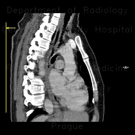
In a patient who ingested lye, CT scans show marked thickening of the esophageal wall due to edema which is more pronounced in the mid and distal part of the esophagus. Note also mild reactive edema in the periesophageal fat, but no signs of perforation.

Tags:  Thorax  Oesophagus
Radiology image - Oesophagitis, corrosive oesophagitis, lye ingestion, CPR: Thorax, Oesophagus: CT - Computed tomography

Oesophagitis, corrosive oesophagitis, lye ingestion, CPR


Oesophagitis, corrosive oesophagitis, lye ingestion, CPR: CT - Computed tomographyOesophagitis, corrosive oesophagitis, lye ingestion: CT - Computed tomographyOesophagitis, corrosive oesophagitis, lye ingestion: CT - Computed tomographyOesophagitis, corrosive oesophagitis, lye ingestion: CT - Computed tomographyOesophagitis, corrosive oesophagitis, lye ingestion, CPR: CT - Computed tomographyOesophagitis, corrosive oesophagitis, lye ingestion, virtual endoscopy: CT - Computed tomographyOesophagitis, corrosive oesophagitis, lye ingestion, virtual endoscopy: CT - Computed tomographyOesophagitis, corrosive oesophagitis, lye ingestion: CT - Computed tomographyOesophagitis, corrosive oesophagitis, lye ingestion: CT - Computed tomography
lights on off