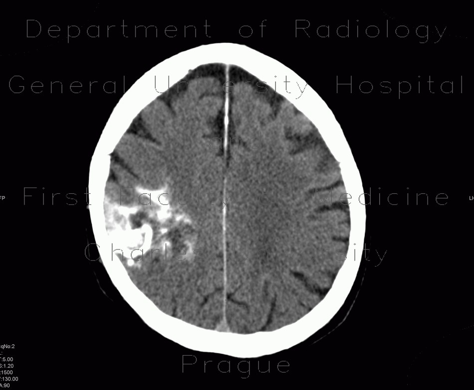
Oligodendroglioma of parietal lobe

CASE

Unenhaced CT scans show typical appearance of a calcified mass in the parietal lobe without signs of perifocal edema. MRI show that the center of the mass contains some fluid, FLAIR sequence show geographic mass of increased signal intensity. The mass causes narrowing of posterior horn of the right lateral ventricle.

Tags:  Brain  Brain
Radiology image - Oligodendroglioma of parietal lobe: Brain, Brain: CT - Computed tomography

Oligodendroglioma of parietal lobe


Oligodendroglioma of parietal lobe: CT - Computed tomographyOligodendroglioma of parietal lobe: CT - Computed tomographyOligodendroglioma of parietal lobe: CT - Computed tomographyOligodendroglioma of parietal lobe: CT - Computed tomographyOligodendroglioma of parietal lobe: CT - Computed tomographyOligodendroglioma of parietal lobe: CT - Computed tomographyOligodendroglioma of parietal lobe: CT - Computed tomographyOligodendroglioma of parietal lobe: CT - Computed tomographyOligodendroglioma of parietal lobe: CT - Computed tomographyOligodendroglioma of parietal lobe: MRI - Magnetic Resonance ImagingOligodendroglioma of parietal lobe: MRI - Magnetic Resonance ImagingOligodendroglioma of parietal lobe: MRI - Magnetic Resonance ImagingOligodendroglioma of parietal lobe: MRI - Magnetic Resonance ImagingOligodendroglioma of parietal lobe: MRI - Magnetic Resonance ImagingOligodendroglioma of parietal lobe: MRI - Magnetic Resonance ImagingOligodendroglioma of parietal lobe: MRI - Magnetic Resonance Imaging
lights on off