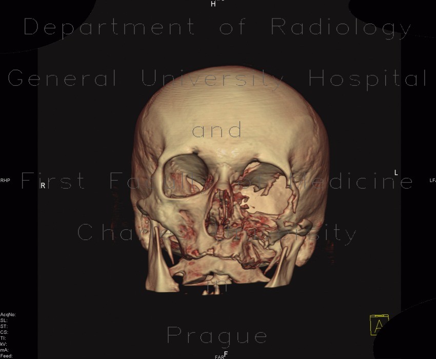
Osteolytic changes of facial skeleton

CASE

Extensive osteolytic destruction of wall of the left maxillary sinus, left half of the sfenoid sinus, medial, lateral wall and floor of the left orbit, and part of the skull base. Osseous structures are replaced by tumorous tissue of nasopharyngeal carcinoma.

Radiology image - Osteolytic changes of facial skeleton: Head and Neck, Bone, Orbit, Sinuses: CT - Computed tomography

Osteolytic changes of facial skeleton


Osteolytic changes of facial skeleton: CT - Computed tomographyOsteolytic changes of facial skeleton: CT - Computed tomographyOsteolytic changes of facial skeleton: CT - Computed tomographyOsteolytic changes of facial skeleton: CT - Computed tomographyOsteolytic changes of facial skeleton: CT - Computed tomographyOsteolytic changes of facial skeleton: CT - Computed tomographyOsteolytic changes of facial skeleton: CT - Computed tomographyOsteolytic changes of facial skeleton: CT - Computed tomographyOsteolytic changes of facial skeleton: CT - Computed tomographyOsteolytic changes of facial skeleton: CT - Computed tomographyOsteolytic changes of facial skeleton: CT - Computed tomographyOsteolytic changes of facial skeleton: CT - Computed tomography
lights on off