ATLAS OF RADIOLOGICAL IMAGES v.1
General University Hospital and 1st Faculty of Medicine of Charles University in Prague
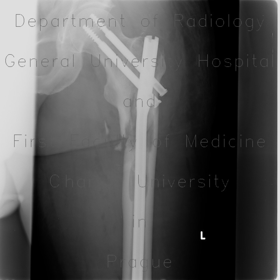
Osteolytic metastasis, pathological fracture of femur, osteosynthesis
CASE
The radiographs show two osteolytic lesions in the proximal femoral shaft and subtrochanteric region, which was a precipitating factor of the subtrochanteric fracture. The fracture was treated with a femoral nail. CT and PET-CT showed several other locations of increased metabolic activity and hypodense osteolytic lesions.


