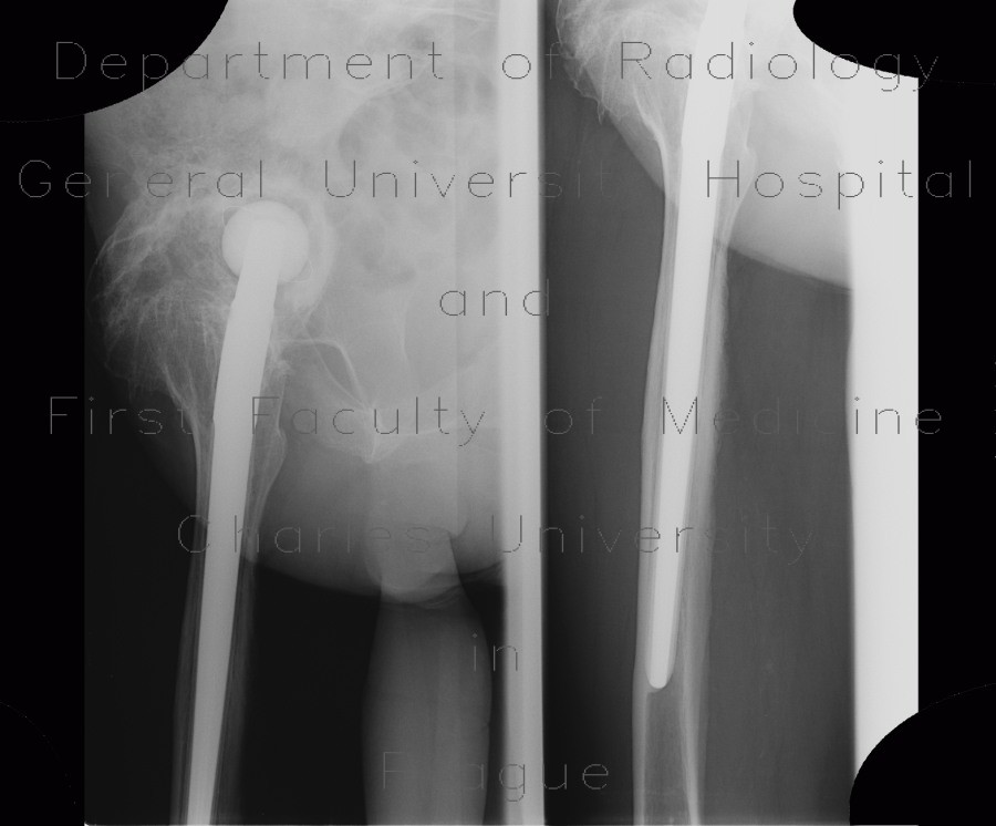
Periarticular ossifications after replacement of hip joint, pseudoarthrosis, false joint

CASE

This patient had both hip joints replaced. He developed massive periarticular calcifications, that were limiting movement in the joint. On the left side a false joint (pseudoarthrosis) developed between the shaft with excessive bone formation and the acetabular rim.

Tags:  Extremity  Bone
Radiology image - Periarticular ossifications after replacement of hip joint, pseudoarthrosis, false joint: Extremity, Bone: X-ray - Plain radiograph

Periarticular ossifications after replacement of hip joint, pseudoarthrosis, false joint


Periarticular ossifications after replacement of hip joint, pseudoarthrosis, false joint: X-ray - Plain radiographPeriarticular ossifications after replacement of hip joint, pseudoarthrosis, false joint: X-ray - Plain radiographPeriarticular ossifications after replacement of hip joint, pseudoarthrosis, false joint: X-ray - Plain radiograph
lights on off