ATLAS OF RADIOLOGICAL IMAGES v.1
General University Hospital and 1st Faculty of Medicine of Charles University in Prague
Pleomorphic adenoma of parotid gland
CASE
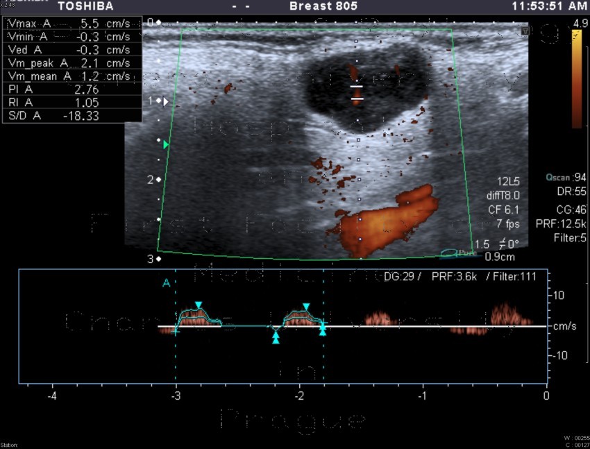
Hypoechoic, well-defined, oval formation with enhanced transmission of ultrasound, thin septae with faint vascularity on color map and high resistance low volume flow on spectral doppler localised in superficial part of the parotid gland. Histology showed pleomorphic adenoma with large mucinous component.


