ATLAS OF RADIOLOGICAL IMAGES v.1
General University Hospital and 1st Faculty of Medicine of Charles University in Prague
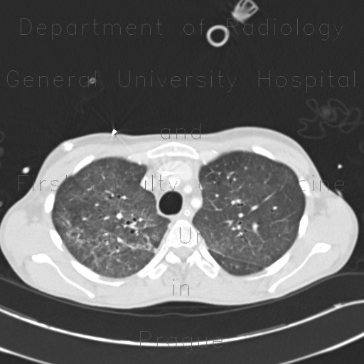
Pneumococcal pneumonia, initial and resolution
CASE
This CT was requested in a imunocompromised patient (leukemia) with new onset of fever. It shows sparse infiltrates mostly of ground-glass density predominantly in the lower lung lobes (dependent position). It was confirmed to be a pneumococcal pneumonia. On a follow-up examination, the infiltrates dissolved leaving inconspicuous areas of faint ground-glass. In general, the first examination was interpreted as atypical pneumonia. Pneumonias caused by streptococcus pneumoniae commonly cause typical pneumonia image with parenchymal consolidation, so this finding is somewhat unusual.


