ATLAS OF RADIOLOGICAL IMAGES v.1
General University Hospital and 1st Faculty of Medicine of Charles University in Prague
Pneumonia
CASE
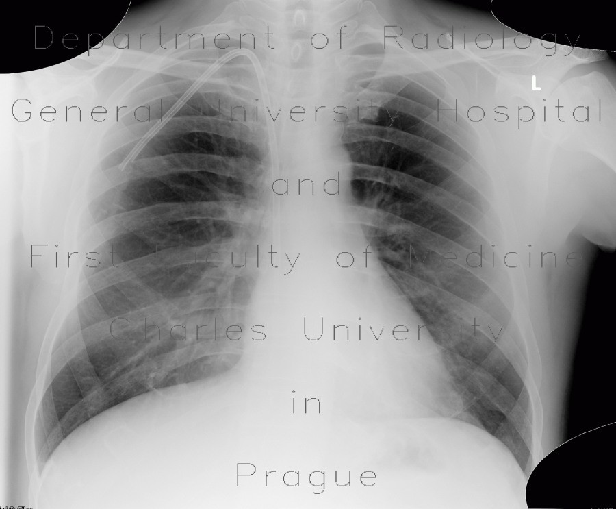
A patient with previously normal chest radiograph developed fever, the cause of which was pneumonia, recognized as consolidation in the left lower and middle lung fields and decreased transparency the left upper lung field. CT showed consolidation of the left lower lung lobe with air-bronchograms.


