ATLAS OF RADIOLOGICAL IMAGES v.1
General University Hospital and 1st Faculty of Medicine of Charles University in Prague
Pneumonia, consolidation
CASE
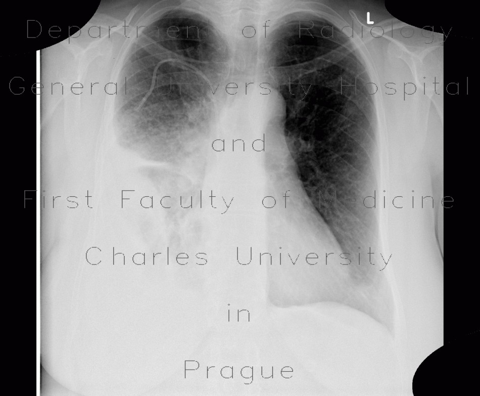
Chest radiograph shows large area of decreased transparency in the right lower to middle lung field which has an apperance of consolidation and loculated pleural fluid. This area is relatively well defined. CT shows large consolidation in the right lower lung lobe and no pleural fluid.


