ATLAS OF RADIOLOGICAL IMAGES v.1
General University Hospital and 1st Faculty of Medicine of Charles University in Prague
Pneumonia, lobectomy
CASE
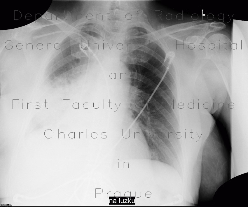
This patient underwent right lower lobectomy and therefore the right hemithorax has decreased volume and the right hemidiaphragm is elevated. Later, he developed pneumonia, which can be seen as an extensive consolidation and ground-glass on CT or air-space shadowing of the right middle and lower lobe.


