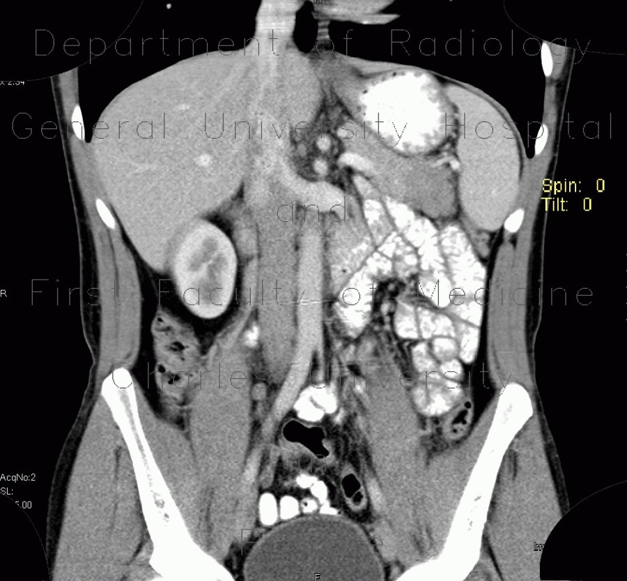
Postinflammatory changes of the tail of pancreas

CASE

CT shows that the tail of pancreas is enlarged and has decreased density. There is no peripancreatic stranding. A differential diagnostis of tumor or postinflammatory changes was stated and MRI was requested. The tail of pancreas has homogeneously increased signal intensity on T2W and decreased on T1 images which indicates postinflammatory changes.

Tags:  Abdomen  Pancreas
Radiology image - Postinflammatory changes of the tail of pancreas: Abdomen, Pancreas: CT - Computed tomography

Postinflammatory changes of the tail of pancreas


Postinflammatory changes of the tail of pancreas: CT - Computed tomographyPostinflammatory changes of the tail of pancreas: CT - Computed tomographyPostinflammatory changes of the tail of pancreas: CT - Computed tomographyPostinflammatory changes of the tail of pancreas: MRI - Magnetic Resonance ImagingPostinflammatory changes of the tail of pancreas: MRI - Magnetic Resonance ImagingPostinflammatory changes of the tail of pancreas: MRI - Magnetic Resonance ImagingPostinflammatory changes of the tail of pancreas: MRI - Magnetic Resonance ImagingPostinflammatory changes of the tail of pancreas: MRI - Magnetic Resonance ImagingPostinflammatory changes of the tail of pancreas: MRI - Magnetic Resonance ImagingPostinflammatory changes of the tail of pancreas: MRI - Magnetic Resonance ImagingPostinflammatory changes of the tail of pancreas: MRI - Magnetic Resonance ImagingPostinflammatory changes of the tail of pancreas: MRI - Magnetic Resonance ImagingPostinflammatory changes of the tail of pancreas: MRI - Magnetic Resonance Imaging
lights on off