ATLAS OF RADIOLOGICAL IMAGES v.1
General University Hospital and 1st Faculty of Medicine of Charles University in Prague
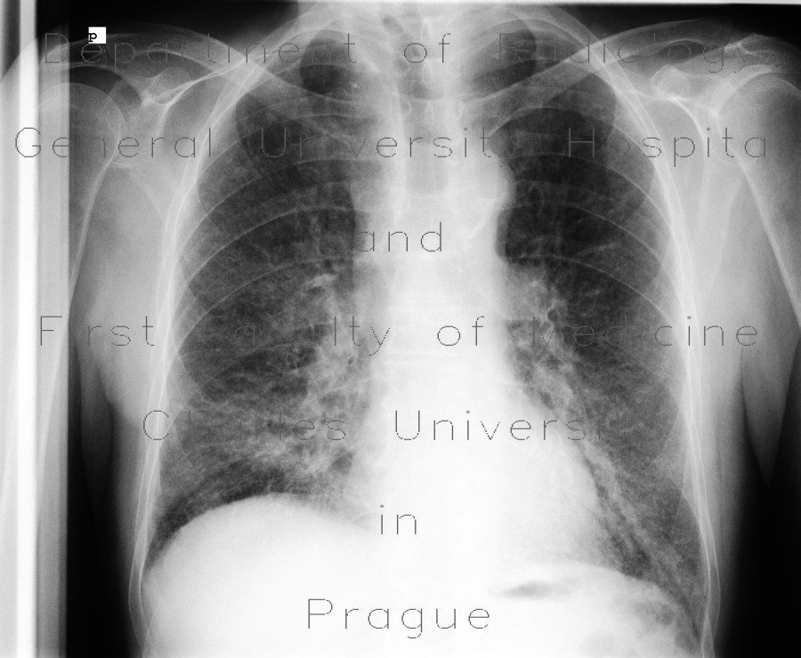
Pulmonary fibrosis, rheumatoid arthritis, alveolitis, exacerbation
CASE
The first chest radiograph shows prominent reticulonodular pattern in both lower and middle lung fields due to fibrosis. The second radiograph shows additional decrease of transparency due to presence of alveolitis as a hallmark of exacerbation of the chronic process.


