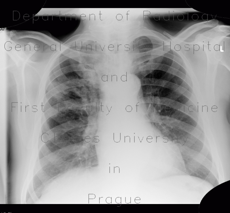
Pulmonary tuberculosis, TBC, tuberculosis

CASE

Both plain chest radiograph and CT show a mass in the right upper lung lobe with a thick walled cavity and extension along bronchovascular bundle in a patient with tuberculosis. Note also right-sided pleural effusion and mild enlargement of mediastinal lymp nodes.

Radiology image - Pulmonary tuberculosis, TBC, tuberculosis: Thorax, Lung, Mediastinum and pleural cavity: X-ray - Plain radiograph

Pulmonary tuberculosis, TBC, tuberculosis


Pulmonary tuberculosis, TBC, tuberculosis: X-ray - Plain radiographPulmonary tuberculosis, TBC, tuberculosis: CT - Computed tomographyPulmonary tuberculosis, TBC, tuberculosis: CT - Computed tomographyPulmonary tuberculosis, TBC, tuberculosis: CT - Computed tomographyPulmonary tuberculosis, TBC, tuberculosis: CT - Computed tomographyPulmonary tuberculosis, TBC, tuberculosis: CT - Computed tomographyPulmonary tuberculosis, TBC, tuberculosis: CT - Computed tomographyPulmonary tuberculosis, TBC, tuberculosis: CT - Computed tomographyPulmonary tuberculosis, TBC, tuberculosis: CT - Computed tomographyPulmonary tuberculosis, TBC, tuberculosis: CT - Computed tomographyPulmonary tuberculosis, TBC, tuberculosis: CT - Computed tomography
lights on off